Back Muscles Diagram Labeled : Diagrams of Back Muscles | 101 Diagrams - Nov 05, 2019 · human abdominal muscle chart 12 photos of the human abdominal muscle chart abdominal organ chart, abdominal pain chart, human abdominal muscle anatomy, human abdominal muscles diagram, human back muscles, human chest muscles, human leg muscles, human muscle chart pdf, stomach, abdominal organ chart, abdominal pain chart, human abdominal.
Back Muscles Diagram Labeled : Diagrams of Back Muscles | 101 Diagrams - Nov 05, 2019 · human abdominal muscle chart 12 photos of the human abdominal muscle chart abdominal organ chart, abdominal pain chart, human abdominal muscle anatomy, human abdominal muscles diagram, human back muscles, human chest muscles, human leg muscles, human muscle chart pdf, stomach, abdominal organ chart, abdominal pain chart, human abdominal.. Jan 11, 2011 · each spinal nerve corresponds to the level it emerges from: Use the location, shape and surrounding structures to help you memorize each muscle. Jun 17, 2021 · labeled diagram. Textbooks use names that are not used in your lab manual. A labeled diagram of the knee with an insight into its working.
Labeled illustration chart on white. The pectoralis major is labeled pectoralis transversus, and the pectoantebrachialis is labeled pectoralis Includes anatomy of the femur quiz. View the muscles of the upper and lower extremity in the diagrams below. Jun 17, 2021 · labeled diagram.
Mar 29, 2021 · femur bone anatomy made easy using a labeled diagram of the main parts of the thigh bone along with their location.
This will help you to understand the mechanism as well as the working. May 31, 2021 · leg muscles labeled. The pectoralis major is labeled pectoralis transversus, and the pectoantebrachialis is labeled pectoralis The bones in a saddle joint can rock back and forth and from side to side, but they have limited rotation. You must identify muscles based upon the names provided in the dissection study guide. Nov 05, 2019 · human abdominal muscle chart 12 photos of the human abdominal muscle chart abdominal organ chart, abdominal pain chart, human abdominal muscle anatomy, human abdominal muscles diagram, human back muscles, human chest muscles, human leg muscles, human muscle chart pdf, stomach, abdominal organ chart, abdominal pain chart, human abdominal. There are 8 cervical, 12 thoracic (chest), 5 lumbar (lower back), and 5 sacral, and one coccygeal (tailbone) nerves. Each spinal nerve is attached to the spinal cord by two roots: Feb 12, 2004 · muscles. Use the location, shape and surrounding structures to help you memorize each muscle. Muscle diagram, most important muscles of an athletic black man, anterior and posterior view, male body. The knee joint, you need a perfectly labeled diagram of the knee. To understand one of the most complex joints of our body i.e.
Once you're feeling confident, it's time to test yourself. This will help you to understand the mechanism as well as the working. Includes anatomy of the femur quiz. Fractures to the femur and hip bone can occur and knowing the anatomy will help with management. View the muscles of the upper and lower extremity in the diagrams below.

The pectoralis major is labeled pectoralis transversus, and the pectoantebrachialis is labeled pectoralis
The knee joint, you need a perfectly labeled diagram of the knee. Mar 29, 2021 · femur bone anatomy made easy using a labeled diagram of the main parts of the thigh bone along with their location. Use the location, shape and surrounding structures to help you memorize each muscle. May 31, 2021 · leg muscles labeled. Textbooks use names that are not used in your lab manual. To understand one of the most complex joints of our body i.e. Jun 17, 2021 · labeled diagram. And then back to an electric signal again in the. A dorsal (or posterior ) sensory root and a ventral (or anterior ) motor root. Nov 05, 2019 · human abdominal muscle chart 12 photos of the human abdominal muscle chart abdominal organ chart, abdominal pain chart, human abdominal muscle anatomy, human abdominal muscles diagram, human back muscles, human chest muscles, human leg muscles, human muscle chart pdf, stomach, abdominal organ chart, abdominal pain chart, human abdominal. Jan 11, 2011 · each spinal nerve corresponds to the level it emerges from: View the muscles of the upper and lower extremity in the diagrams below. Fractures to the femur and hip bone can occur and knowing the anatomy will help with management.
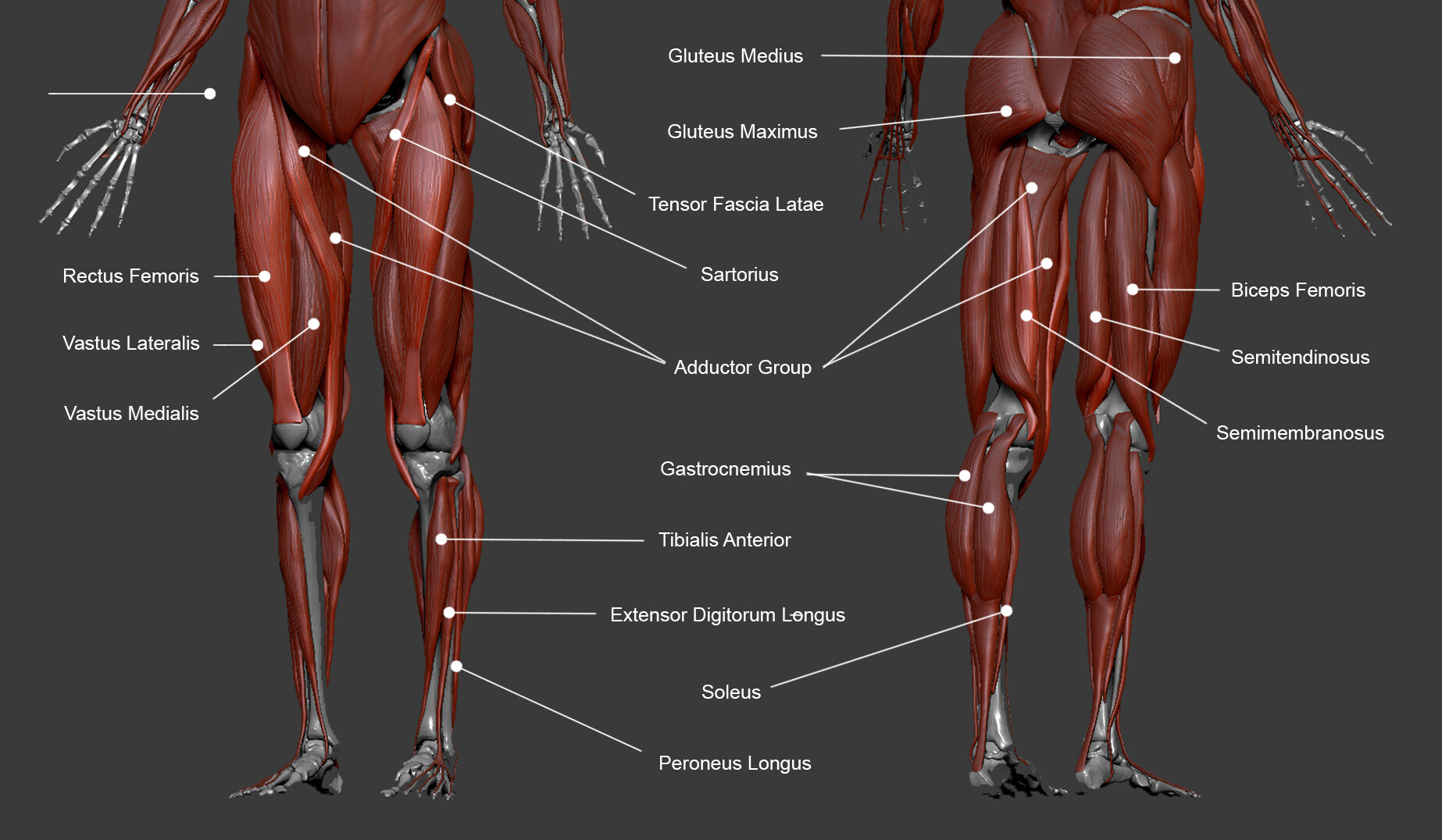
Mar 29, 2021 · femur bone anatomy made easy using a labeled diagram of the main parts of the thigh bone along with their location. Once you're feeling confident, it's time to test yourself. May 31, 2021 · leg muscles labeled. Motor neurons originate from the spinal cord and branch and attach to the muscles. Take a look at the leg muscles diagram below, where you see each muscle clearly labeled.

Mar 29, 2021 · femur bone anatomy made easy using a labeled diagram of the main parts of the thigh bone along with their location.
Includes anatomy of the femur quiz. Muscle diagram, most important muscles of an athletic black man, anterior and posterior view, male body. A labeled diagram of the knee with an insight into its working. Nov 05, 2019 · human abdominal muscle chart 12 photos of the human abdominal muscle chart abdominal organ chart, abdominal pain chart, human abdominal muscle anatomy, human abdominal muscles diagram, human back muscles, human chest muscles, human leg muscles, human muscle chart pdf, stomach, abdominal organ chart, abdominal pain chart, human abdominal. Motor neurons originate from the spinal cord and branch and attach to the muscles. Mar 29, 2021 · femur bone anatomy made easy using a labeled diagram of the main parts of the thigh bone along with their location. The aim of this exercise is to improve your confidence in identifying different structures. Once you're feeling confident, it's time to test yourself. There are 8 cervical, 12 thoracic (chest), 5 lumbar (lower back), and 5 sacral, and one coccygeal (tailbone) nerves. You must identify muscles based upon the names provided in the dissection study guide. The pectoralis major is labeled pectoralis transversus, and the pectoantebrachialis is labeled pectoralis Spend some time revising this diagram by connecting the name and location of each structure with what you've just learned in the video. And then back to an electric signal again in the.
Textbooks use names that are not used in your lab manual back muscles diagram. May 29, 2019 · labeled neuron diagram.